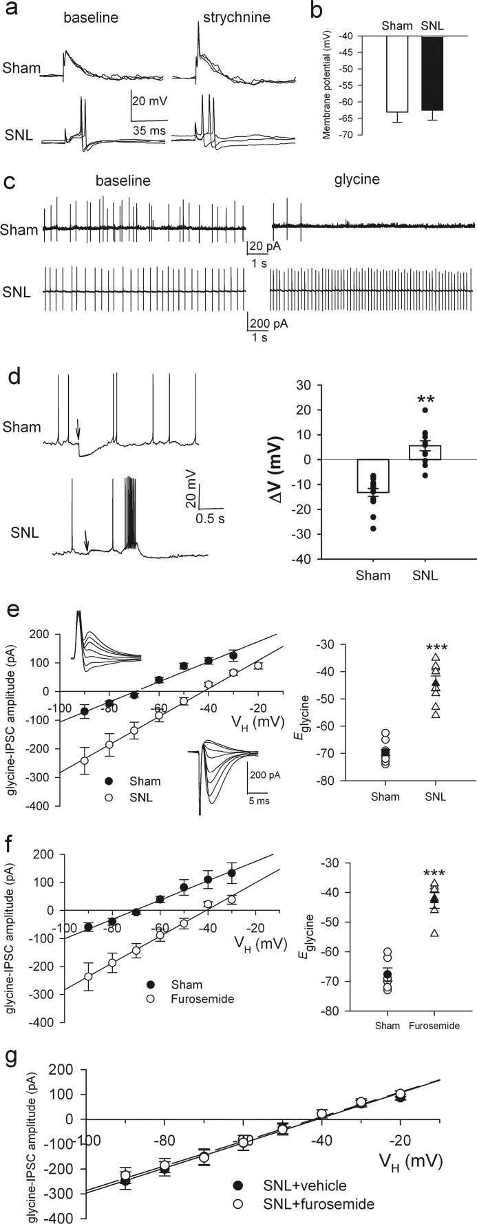
FIGURE 1.
Nerve injury causes diminished glycine-mediated synaptic inhibition, a depolarizing shift in Eglycine of lamina II neurons, and KCC2 down-regulation in the spinal cord. a, perforated recordings show EPSP-spike activity of lamina II neurons evoked by electrical stimulation of the dorsal root in sham control (top) and SNL (bottom) rats before and during bath application of 2 μm strychnine. Responses evoked by three consecutive stimulations (at 15-s intervals) were superimposed. b, mean resting membrane potentials of lamina II neurons in sham control (n = 15) and SNL (n = 15) rats (p = 0.92, Mann-Whitney test) are shown. c, cell-attached recordings show the spontaneous firing activity of lamina II neurons from sham control (top) and SNL (bottom) rats before and during bath application of 1 mm glycine. d, perforated recordings (left) and mean membrane potential changes (right) induced by puff application of 300 μm glycine to lamina II neurons in sham control (n = 15) and SNL (n = 12) rats are shown. The downward arrows indicate the time of puff glycine application. e, I-V plots show glycine-mediated IPSCs (left) and mean changes in Eglycine (right) in lamina II neurons from sham control (n = 10) and SNL (n = 8) rats. Inset, original traces of glycine-mediated IPSCs recorded using perforated patch-clamp at different holding potentials (VH) from −90 to +30 mV at 20-mV steps. f, I-V plots show glycine-mediated IPSCs (left) and changes in Eglycine (right) in lamina II neurons (n = 6) of control rats before and during bath application of 200 μm furosemide. g, I-V plots show Eglycine of lamina II neurons in spinal cord slices of SNL rats treated with furosemide (n = 6) or vehicle (n = 7). VH, holding potential. **, p < 0.01; ***, p < 0.001, Mann-Whitney test. Error bars represent S.E.

