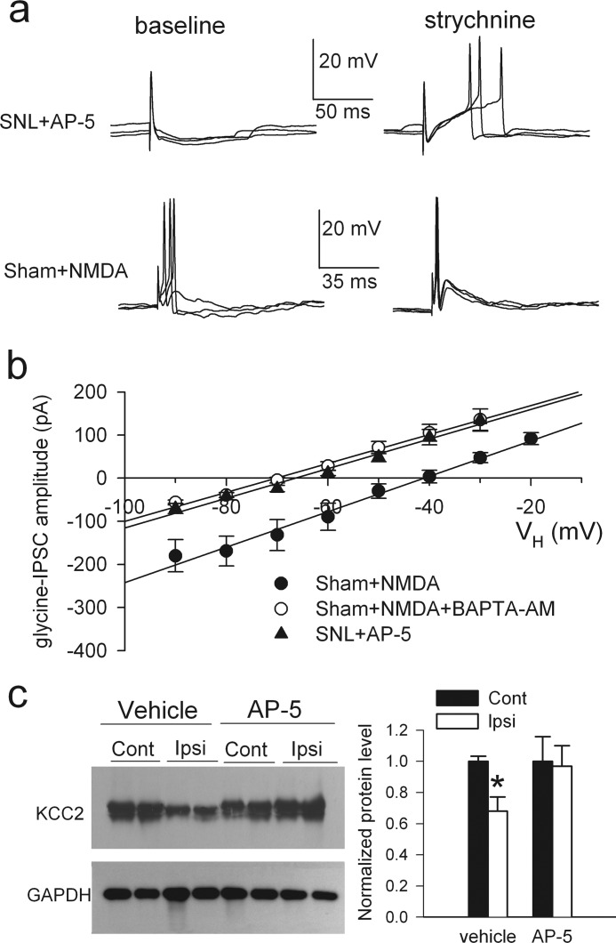
FIGURE 3.
Nerve injury diminishes synaptic inhibition and the depolarizing shift in Eglycine of lamina II neurons through NMDAR activation. a, perforated recordings show the effects of bath application of 2 μm strychnine on EPSP-spike activity of lamina II neurons evoked by dorsal root stimulation in slices taken from control rats incubated with NMDA (30 μm, 2.5–3 h) or in slices taken from SNL rats incubated with AP-5 (50 μm, 3 h). b, I-V plots show changes in Eglycine of lamina II neurons from slices of control rats incubated with NMDA alone (n = 7) or NMDA plus BAPTA-AM (50 μm, n = 6) and from slices taken from SNL rats incubated with AP-5 (n = 7). VH, holding potential. c) shown are Western blots (left) and quantification (right) of KCC2 protein (∼140 kDa) amounts in the dorsal spinal cords ipsilateral (Ipsi) and contralateral (Cont) to SNL in rats treated with intrathecal administration of AP-5 (n = 7) or vehicle (saline, n = 8). KCC2 was detected by using KCC2 N terminus antibody. * p < 0.05, Kruskal-Wallis test. Error bars represent S.E.

