
FIG. 2.
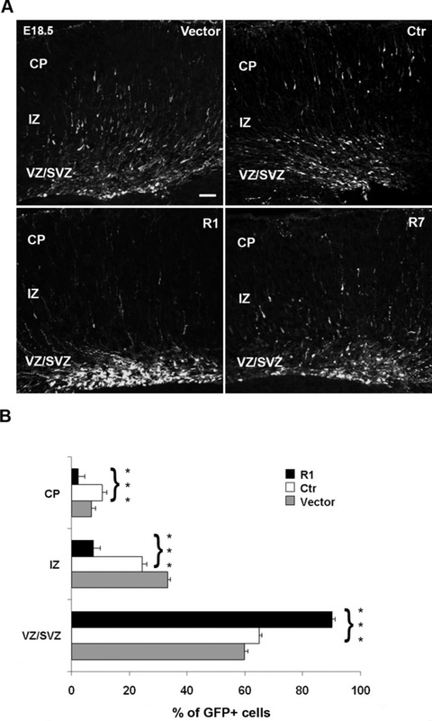
Effect of α5 integrin knockdown in developing murine cerebral cortex. (A) Representative coronal sections of embryonic murine neocortex 3 days (E18.5) following electroporation of empty vector, Ctr, R1 and R7 α5 shRNAs together with coral GFP. Transfection of R1 and R7 shRNAs impaired radial migration. Scale bar, 100 μm. (B) Quantitative analyses of the distribution of GFP-positive cells in the various cortical layers. The cortical wall was subdivided, and numbers of GFP-positive cells were counted in each layer and expressed as a percentage of the total. Statistical differences were seen for each layer, comparing the distribution of GFP-positive cells between Ctr and R1 α5-shRNA. Asterisks indicate significant differences between the groups (***P < 0.001, using Student’s t-test).