
Figure 3. Jab1 specifically interacts with p27.
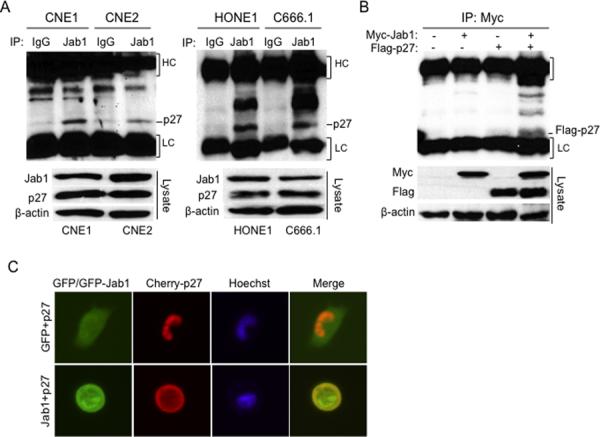
(A) Cell lysates were immunoprecipitated with either non-immune mouse serum or Jab1 and immunoblotted with anti-p27. Immunoglobulin G heavy chain (HC) and light chain (LC) were indicated. (B) 293T cells were transfected with either Myc-Jab1 or Flag-p27 or both for 48 hours, and then cell lysates were immunoprecipitated with Myc and immunoblotted with Flag. Cell lysates immunoblotted with the indicated antibodies are shown in the bottom panel. (C) CNE1 cells were transfected with a GFP vector and Cherry-p27 or GFP-Jab1 and Cherry-p27 for 48 hours, and then examined under a fluorescence microscope. Original magnification ×200.