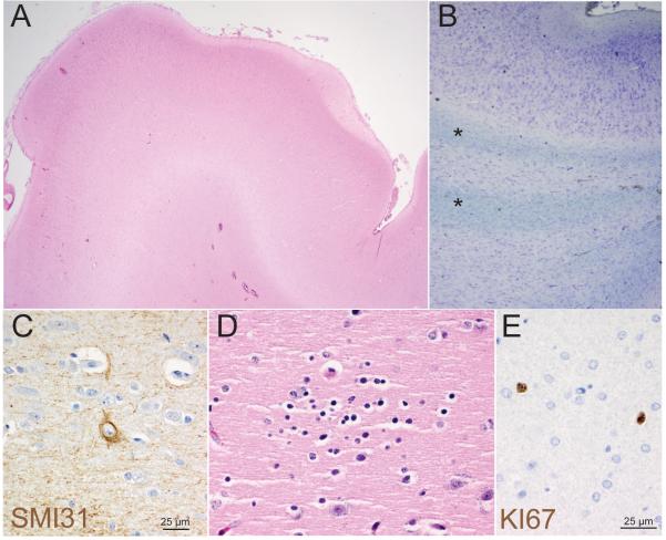
Figure 2. Abnormal cortical development in hemimegalencephaly case HMG-1 with trisomy of chromosome 1q.
(A) Low-power view (20x mag) of a gyrus from the cerebral cortex stained with haematoxylin and eosin (H&E) shows an abnormally contoured surface and variably thick cortical ribbon and molecular layer. (B) Analysis of subcortical white matter using Cresyl violet and Luxol Fast Blue (LFB) highlights numerous subcortical bands/islands of ectopic gray matter containing neurons and glia (*). (C) Immunohistochemical staining for phosphorylated neurofilament, SMI31, highlights scattered abnormal large neurons. (D) Rare small collections of neuroblast-like cells (microdysplasia) were present on H&E. (E) Immunohistochemical staining also demonstrated an abnormal number of proliferating Ki67 positive cells scattered throughout gray and white matter which had an atypical nuclear morphology (e). (A) 20x, (B) 200x (C, D, E) 600x.

