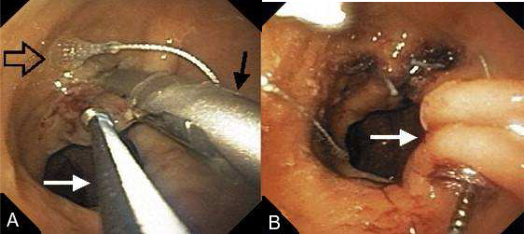
FIGURE 12.
(A) After tissue is grasped at rim of the GJA with a corkscrew-like tissue grasper (white arrow), the g-Prox is closed on tented tissue (black arrow) and the first anchor is deployed (open arrow). (B) after the g-Prox is released from the tissue, the second anchor is deployed, creating a tissue plication (arrow). (Permission to reuse image obtained from Elsevier)

