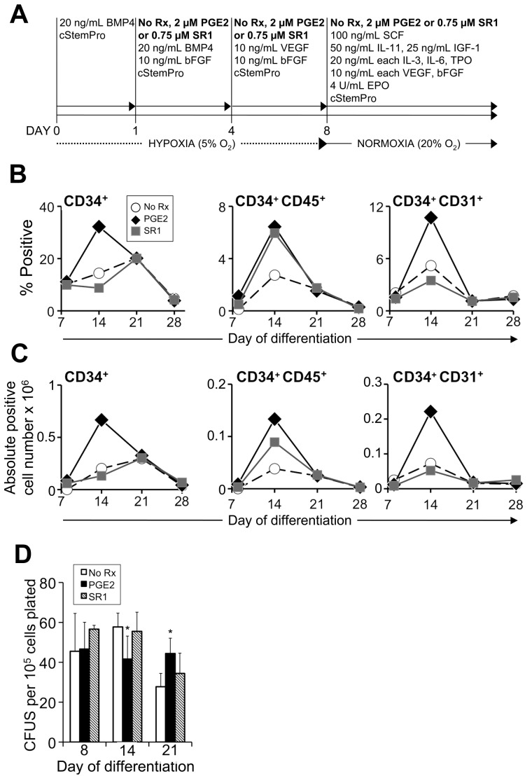
Figure 2.
PGE2 and SR1 alter the kinetics and efficiency of hematopoietic differentiation of MniPSCs. (A) Schematic of hematopoietic differentiation with or without the addition of PGE2 or SR1. On day 0, MniPSC line 3 colonies (passage 50) were aggregated into EBs in cStemPro medium with BMP4. On day 1, media were supplemented with 2μM PGE2, 0.75μM SR1, or untreated (No Rx). For each subsequent induction and media change until the end of the experiment, cStemPro was supplemented with PGE2 or SR1. (B) Comparative kinetics of hematopoietic specification for PGE2, SR1, or untreated cultures as determined by flow cytometry analysis. On the indicated days of differentiation, EBs were dissociated, counted, stained with fluorophore-conjugated antibodies and analyzed by flow cytometry. (C) Absolute cell number of fluorphore+ cells. Absolute cell yields corresponding to the indicated hematoendothelial subsets were calculated (absolute positive cell yield = 100 × [% fluorphore+ × viable cell yield]). Each number shown is ×106 and is representative of the total fluorphore+ cell yield obtained from an input undifferentiated MniPS cell number of 1 million. (D) Hematopoietic colony-forming cell assays plated on the indicated days of differentiation. MniPSC-derived cells were dissociated and single cells plated in Methocult H4435. CFUs were enumerated and scored as a function of input cell number (CFUs per 105 cells plated). The results from 1 representative experiment of 3 conducted are shown. Error bars indicate SD of mean of triplicates. ANOVA significance levels: *P = .042 (day 14), *P = .039 (day 21).

