Figure 3.
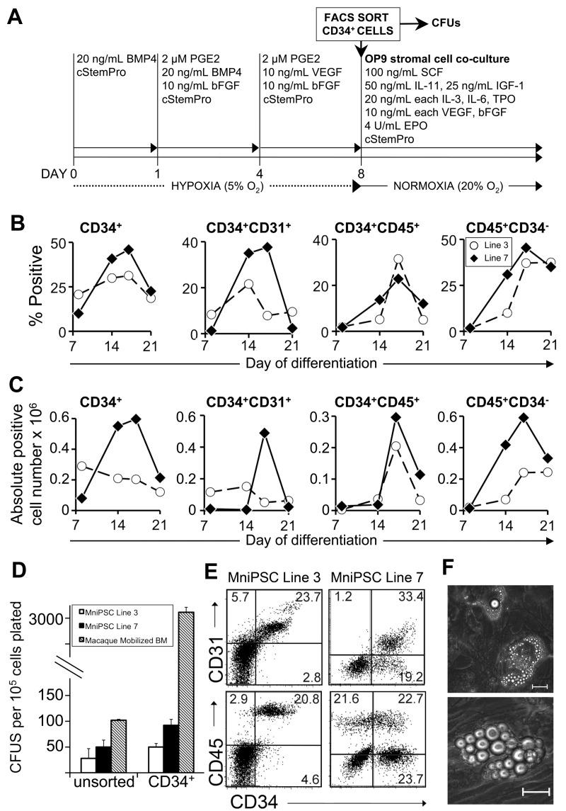
FACS-sorted MniPSC-CD34+ cells give rise to CD45+CD34− hematopoietic progeny. (A) Schematic of differentiation of MniPSC lines 3 and 7. Both MniPSC lines were induced days 0 to 8 with the optimized protocol outlined in Figure 2 with 2μM PGE2 added to EBs on days 1-8 of differentiation. On day 8, EBs were dissociated into single cells, immunostained with PE-conjugated anti-CD34 antibody (clone 563), and sorted by FACS for the CD34high fraction. CD34high cells were plated in cStemPro plus cytokines (50 000 CD34high cells per well of a 6-well plate) on irradiated OP9 stromal cells. (B) Percentages of CD34high cells that coexpress hematoendothelial markers on the indicated days of differentiation, which include presorted (day 8) and postsorted populations (days 14, 21). (C) Absolute number of fluorphore+ cells for MniPSC lines 3 and 7 that were induced toward hematopoietic differentiation, sorted for the CD34high fraction on day 8, and plated in OP9 coculture. Absolute cell yields corresponding to the indicated hematoendothelial subsets were calculated (absolute positive cell yield = 100 × [% fluorphore+ × viable cell yield]). Each number shown is ×106 and is representative of the total fluorphore+ cell yield obtained from an input undifferentiated MniPS cell number of 1 million. (D) Comparison of hematopoietic colony-forming potential (CFUs) of unsorted and CD34-enriched cells from day 8 MniPSC line 3 and line 7 HPCs and pigtail macaque mobilized bone marrow. To evaluate MniPSC-derived HPCs, EBs were dissociated on day 8, kept in bulk (unsorted), or sorted by FACS and then plated in Methocult H4435. Error bars indicate SD of mean of 3 separate experiments. (E) Representative flow cytometry dot plots of MniPSC-derived HPCs on day 18 of differentiation. (F) Representative images of MniPSC Line 7-derived CD34high cell-derived hematopoietic zones on OP9 stromal cells on day 21 of differentiation. Similar results were obtained from MniPSC line 3 CD34high cells. Colonies were imaged on a Nikon Eclipse Ti-M (TiSR) microscope and photographed with a Nikon camera, in cStemPro media (10× [left] and 20× [right] objectives). Scale bar = 100 μm.

