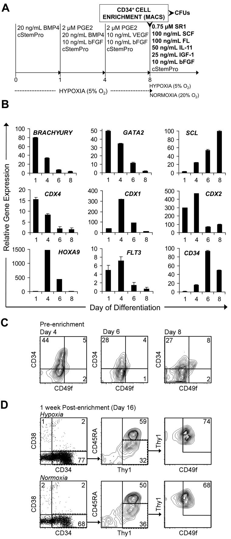
Figure 4.
SR1 maintains and expands MniPSC-derived CD34+ HPCs purified by MACS enrichment. (A) Schematic of MniPSC HPC cell differentiation, enrichment, and expansion. MniPSC Line 7 (Passage 54) cells were aggregated and induced toward differentiation as shown in Figure 3. On day 9 of induction, cells were purified by MACS with anti–human CD34 antibody (clone 12.8). The CD34+ fraction was then cultured under hypoxic (5% O2) or normoxic (20% O2) conditions in cStemPro containing the indicated cytokines and 0.75μM SR1 for HPC expansion. (B) qRT-PCR–based expression of hematopoietic specific genes on days 1, 4, 6, and 8 of hematoendothelial differentiation. Expression levels are relative to β-actin and calibrated to undifferentiated MniPSCs. Error bars indicate SD of mean of triplicate samples from 1 representative experiment of 3. (C) Flow cytometry analysis of CD34 and CD49f coexpression on days 4, 6, and 8 of differentiation before enrichment by MACS. (D) Flow cytometry analysis of putative LT-HSCs (CD34+CD38−Thy1+CD45RA−CD49f+) after a 1-week expansion in SR1 under hypoxic (top) or normoxic (bottom) conditions. Fold expansion of total CD34+CD38−Thy1+CD45RA−CD49f+ cells: 1.1 (hypoxia), 3 (normoxia).

