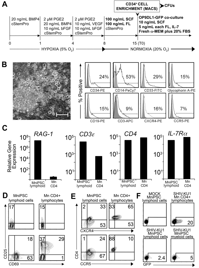
Figure 5.
MniPSC-derived CD34+ HPCs exhibit myeloid and lymphoid differentiation potential. (A) Schematic of differentiation. MniPSC Line 7 cells (Passage 18) were induced according to the optimize protocol outlined in the previous figure schema. On day 8, EBs were replated in cStemPro plus SCF and FL for HPC expansion. On day 15, CD34+ cells were MACS purified and plated into Methocult (myeloid CFU assays) or into stromal cell coculture with OP9 cells that express GFP and the Notch ligand Delta-Like 1 (OP9DL1-GFP). To induce lymphoid differentiation, cells were cultured in freshly prepared α-MEM containing FBS, SCF, FL, and IL-7. T0 indicates the starting time point for lymphocyte differentiation. (B) Myeloid phenotype of MniPSC-derived hematopoietic progeny. Left, Representative CFU-M colony differentiated from day 14 CD34+ HPC cells. Colonies were imaged on a Nikon Eclipse Ti-M (TiSR) microscope and photographed with a Nikon camera, in Methocult (20× objective). Scale bar = 100 μm. Right, Flow cytometry analysis of myelomonocytic hematopoietic colonies isolated from Methocult. Myelomonocytic colonies from day 14 MniPSC line 7 CD34high HPCs were isolated from Methocult after CFU scoring on day 24 and analyzed. Gray histograms indicate isotype controls for the indicated fluorophores. (C) qRT-PCR–based expression analysis of T lymphocyte–specific genes in MniPSC-derived lymphoid cocultures (day 30 of lymphoid differentiation). Gene expression also was assessed in CD4+ lymphocytes purified from macaque peripheral blood mononuclear cells and corrected for OP9DL1 stromal cell gene expression. Gene expression levels are relative to β-actin and calibrated to levels detected in undifferentiated MniPSCs. Error bars indicated the SD of the mean of quadruplicate samples. (D) Up-regulation of early activation markers in MniPSC lymphoid cells. Macaque (Mn) CD4+ lymphocytes and day 45 MniPSC lymphoid cultures were untreated or activated for 24 hours with hIL-2 and CD3/CD28 beads and then analyzed by flow cytometry. (E) Expression of HIV entry coreceptors in activated macaque (Mn) CD4+ lymphocytes and day 45 MniPSC-derived lymphoid cultures. (F) MniPSC-derived myeloid and lymphoid cells support SHIVKU-1 entry and viral replication. GFP expression in Ghost cells cultured with supernatant from CD4+ lymphocytes, MniPSC myeloid and lymphoid cultures infected with SHIV-KU1 after virus washout.

