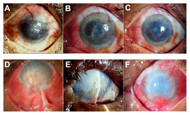
Figure 4.
Slit-lamp photographs show a comparison of the eyes before (left column) and after (middle and right column) cultivated limbal epithelial transplantation (CLET) in the histopathologic failure group. Patient 10 (top row) has total limbal stem cell deficiency from a severe chemical burn (A). Postoperatively, the patient has histopathologic failure but clinical success. Cytology shows goblet cells in the central cornea at 12 months, but the central cornea has remained clear with minimal haze and no vascularization. The visual acuity at 13 (B) and 29 (C) months remains 20/125. Patient 9 (bottom row), with limbal stem cell deficiency from a severe chemical burn, has marked lid abnormality and corneal stromal destruction (D). The patient undergoes auto-CLET. Seven months postoperatively (E), the ocular surface has less inflammation and no recurrent symblepharon. However, an epithelial defect persists and does not heal after subsequent tarsorrhaphy surgery. The patient finally undergoes penetrating keratoplasty at 9 months. Complete epithelialization has not been achieved and the clinical course was complicated by secondary infectious keratitis. The corneal graft is then rejected and eventually opaque with complete epithelialization at 30 months (F).
Note: A bandage contact lens is applied to protect against mechanical trauma from the lid and eyelashes.

