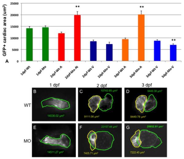
Fig. 5. Chamber sizes are altered in the tbx2ab morphants.
Hearts were imaged in wildtype or tbx2ab double morphant embryos and the area of atrial and ventricular chambers was outlined and measured. The top panel (A) shows the quantification from representative embryos at 1 dpf, 2 dpf, or 3 dpf as indicated. Also as indicated, the measurement was in wildtype (WT) or morphant (MO), either in the heart tube (1 dpf, green), or for the atrium (A) or ventricle (V). In each case n is at least 10. Lower panels show representative images of wildtype and tbx2ab morphant hearts at 1 dpf (B, E), 2 dpf (C, F) and 3 dpf (D, G). In B and E the heart tube is outlined in green. In C, D, F, and G, yellow outlines the ventricle and green outlines the atrium. The ** indicates statistical significance compared to wildtype, according to Student’s t-test, p<0.01.

