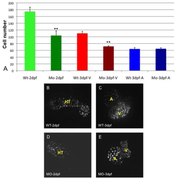
Fig. 6. The number of cardiomyocytes in tbx2ab morphants compared to wildtype is not different in the expanded atrium, but is relatively decreased in smaller ventricles.
Hearts were imaged using embryos from the myl7:dsRed-nuc reporter line and numbers of cardiomyocytes counted manually in Z-stack sections. The top panel (A) shows the quantification of data for wildtype (WT) or double morphant embryos (Mo) at 2 dpf or 3 dpf as indicated. Also as indicated, the cells were counted in the heart tube (HT) at 2 dpf or specifically in the ventricle (V) or atrium (A) at 3 dpf. The ** indicates statistical significance compared to wildtype, according to Student’s t-test, p<0.01. Lower panels show representative images of wildtype (WT) or tbx2ab morphants (MO) at 2 dpf (B, D) and 3 dpf (C, E). For each measurement n is at least 10.

