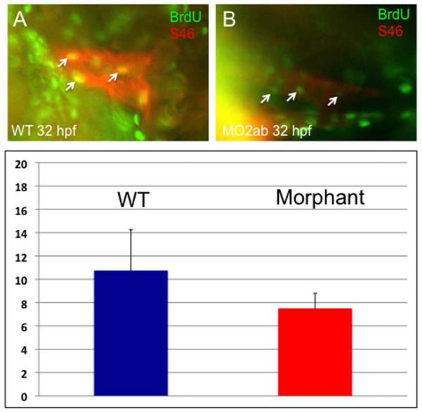
Fig. 9. Cardiomyocyte proliferation is not decreased in the atrium of tbx2ab morphants.
Embryos were pulse-labeled with BrdU, and after being fixed, were co-stained to detect BrdU+ cells (green) and S46+ atrial cardiomyocytes (red). Hearts were imaged and the yellow (double positive) cells counted (examples indicated by the small arrows). The top panels show representative wildtype (A) and morphant (B) embryos at ~32 hpf. The lower panel (C) shows quantification of the average BrdU+ cardiomyocytes, in each case from several randomly chosen embryos, n = 4. According to Student’s t-test, p=0.13. The result is consistent with the fact that the atrium is not altered in cell numbers.

