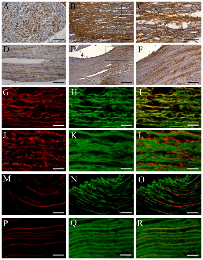
Figure 3. Localization of AQP1 in the peripheral and central branches of human (A–C,G–L) and mouse (D–F,M–S) trigeminal neurons.
(A–F) Representative images of immunohistochemistry for AQP1. (G–R) Representative images of double immunofluorescence with AQP1 (red) and GFAP (green in H and N) or β-tubulin III (green in K and Q). A considerable proportion of axons are positive for AQP1 within the human (transverse section, A) and mouse (longitude section, D) mandibular nerve. The human trigeminal central root exhibits dense immunoreactivity for AQP1 (B, C, G, J), which is colocalized with GFAP (I), but not β-tubulin III (L). In contrast, in the mouse trigeminal central root, there are a population of AQP1 positive axons (E, F, M, P) that coexpress β-tubulin III (R) but not GFAP (O). Scale bars = 100 µm in A, D, F; 300 µm in B; 200 µm in E; 50 µm in C, G–R.

