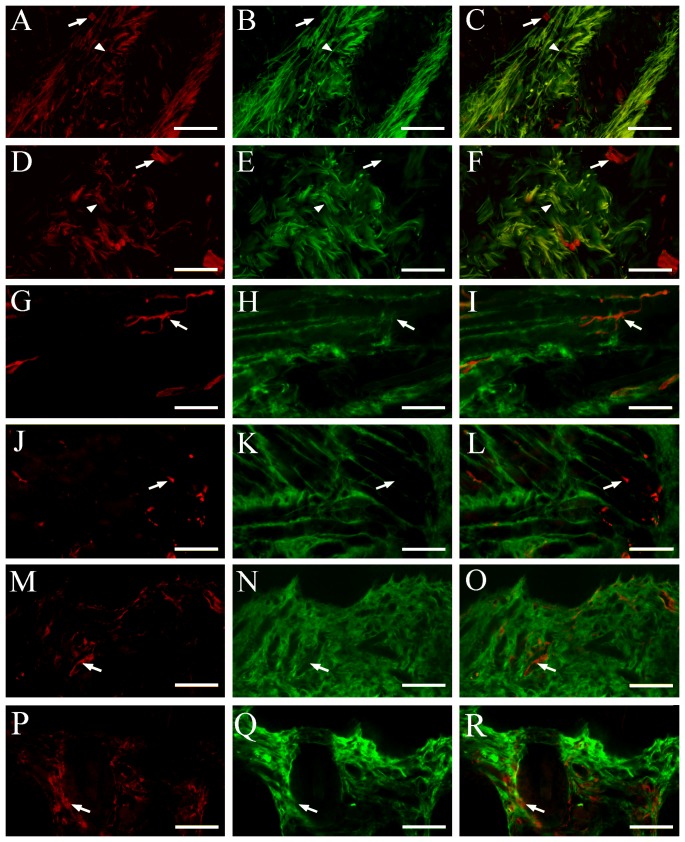
Figure 6. Colocalization of AQP1 and β-tubulin III in the oral submucosa, muscle and skin of humans (A–C, G–I, M–O) and mice (D–F, J–L, P–R).
(A–F) AQP1 (red) and β-tubulin III (green) are coexpressed within a number of the nerve bundles (arrowheads) of the cheek submucosa from the two species. AQP1 positive microvessels (arrows) are also scatted among the nerve bundles. (G–R) There are a large number of β-tubulin III (green) positive nerve fibers, and a few AQP1 positive microvessels (arrowheads), within the cheek intermusclar (G–L) and subcutaneous regions (M–R). No overlap between β-tubulin III and AQP1 immunoreactivity is observed. Scale bars = 100 µm.

