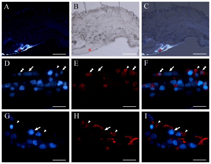
Figure 7. Mouse AQP1 positive trigeminal neurons innervate the oral mucosa but not skin.
(A–C) Representative combined fluorescent and light images showing the injection site of retrograde tracer FG (red asterisks) within the cheek mucosa and submucosa of mice. (D–I) Representative double fluorescent images showing the co-distribution of AQP1 immunoreactive neurons (blue) with FG-labeled neurons (green) innervating cheek mucosa (D–F) or skin (G–I). AQP1 and FG double-labeled neurons are marked with arrowheads. AQP1 single-labeled neurons are marked with the double-arrowheads, and FG single-labeled neurons with arrows. Note that some FG-labeled small-size mucosal neurons are co-labeled with AQP1. No FG-labeled cutaneous neurons are positive for AQP1. Scale bars = 500 µm in A–C; 40 µm in D–I.

