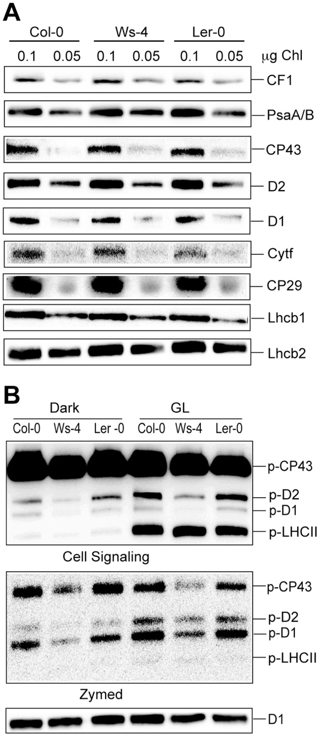
Figure 6. Levels of thylakoid photosynthetic proteins in Col-0, Ws-4 and Ler-0 accessions.

Thylakoid membranes were isolated from plants grown hydroponically at an irradiance of 120 µmol photons m−2 s−1 (GL). (A) Representative western blots with antibodies against various photosynthetic proteins. The loaded amount of Chl (µg) is indicated above each lane. (B) Phosphorylation of PSII proteins was assessed in thylakoid membranes isolated in the presence of 10 mM NaF from plants dark-adapted for 16 h or exposed for 3 h to GL. Representative western blots with anti-phosphothreonine antibody from Cell Signaling and Zymed and with anti-D1 antibody (as loading control) are shown. The gels were loaded with 0.25 µg of Chl in each well. The positions of the major phosphorylated PSII proteins are indicated.
