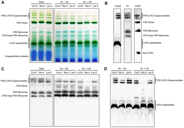
Figure 11. Analysis by Blue-native gel electrophoresis of thylakoid protein complexes from Col-0, Ws-4 and Ler-0 accessions.
Leaves detached from plants grown hydroponically at an irradiance of 120 µmol photons m−2 s−1 (GL) were exposed to high light (HL = 950 µmol photons m−2 s−1) in the absence or presence of lincomycin (LN) for 3 h. As control, 16 h dark-adapted plants were used. Thylakoid membranes were isolated and solubilized mildly with n-dodecyl-ß-D-maltoside, and the various types of Chl protein complexes were separated by Blue-native gel electrophoresis. (A) Representative unstained Blue-native gel (8 µg Chl per lane). (B) (C). The PSII complexes together with various combinations of LHCs were identified based on western bloting with anti-Lhcb2, D1 and CP43 antibodies, and based on previous reports [2], [33]. Representative western blot with anti-D1 antibody of gel as in (A). (D) Representative western blot with anti-Lchb2 of gel as as in (A).

