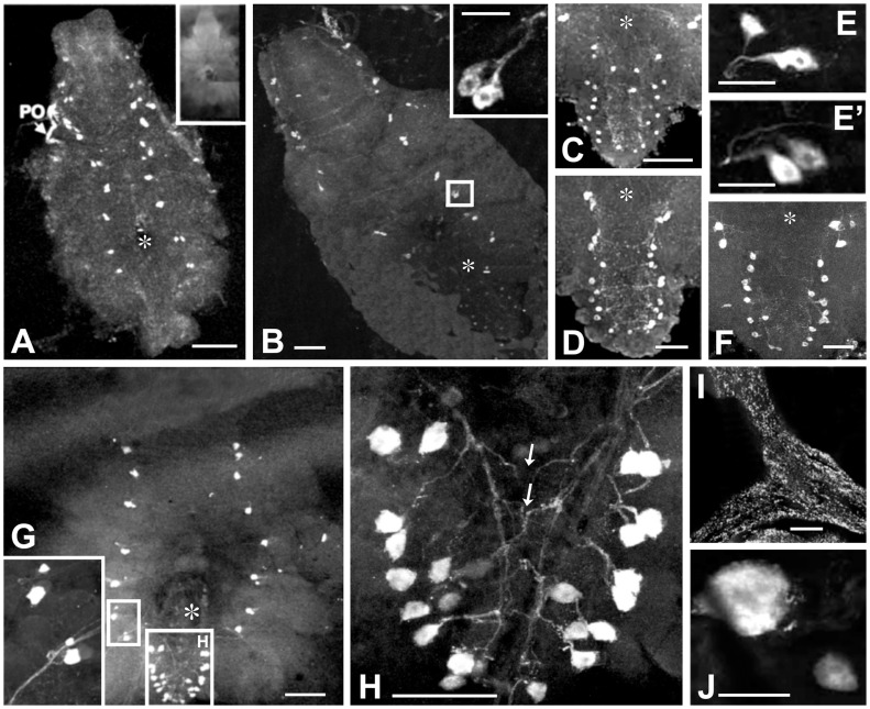
Figure 2. Immunolocalization of two different types of bilaterally paired CasBurs neurons per neuromere in crab TGC and POs during late postembryonic development.
(A) CasBurs neurons in the subesophageal and thoracic but not in the abdominal neuromeres of TGC of a postlarva, megalopa. Inset shows a pre-absorption control of an adult TGC. (B) CasBurs neurons in the entire TGC of the 1st crab. (C–F) Details of neurons in the last thoracic and abdominal neuromeres of the 1st(C), 3rd (D), 4th crab (F). (E, E’) Two distinct types of CasBurs neurons in thoracic segments showing a difference in staining intensity but no size disparity yet. (G) CasBurs neurons in the TGC of a 12th crab, inset: enlarged cells in the 4th and the 5th thoracic neuromeres; note clear disparity in size of the distinct neuron types in abdominal neuromers. (H) Higher magnification of abdominal neuromeres of the 12th crab’s TGC without apparent size disparity; note the contralateral projection patterns (arrows). (I) Fibers and terminals in the anterior bar of a PO (J) A pair of typically disparate-sized thoracic cell bodies in an adult TGC. Asterisks: foramen of the sternal artery. Whole mounts in all cases; scale bars A, B, C, D, F, I = 100 µm; B inset, E, E’ = 50 µm; H = 200 µm; J = 25 µm.

