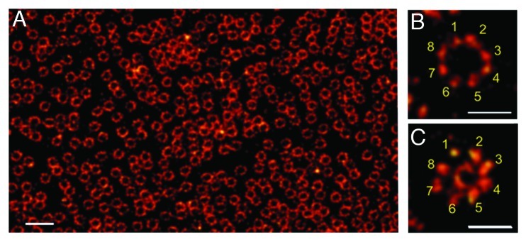
Figure 2. Nuclear envelope of a Xenopus laevis oocyte as seen by dSTORM.33 (A) Nuclear envelopes isolated from Xenopus laevis oocytes were labeled with Alexa647 by indirect immunofluorescence against gp210, a protein that localizes to the lumen of the nuclear envelope bordering the pore wall. (B) Higher magnifications reveal the structural arrangement of gp210 proteins in nuclear pore complexes (NPCs). The 8-fold symmetry of the gp210 ring around each NPC (B) and the diameter of the central channel of ~40 nm is correctly identified with an optical resolution of ~15 nm (C) using WGA-Alexa647 binding to nucleoporins of the central channel (ref. 33). Scale bars: 500 nm (A), 150 nm (B, C).

An official website of the United States government
Here's how you know
Official websites use .gov
A
.gov website belongs to an official
government organization in the United States.
Secure .gov websites use HTTPS
A lock (
) or https:// means you've safely
connected to the .gov website. Share sensitive
information only on official, secure websites.
