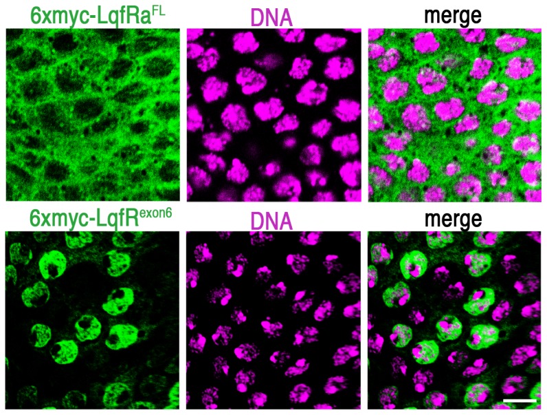
Figure 3. Subcellular localization of Myc-tagged LqfR proteins.

Confocal microscope images of third instar larval eye disc tissue from two different discs (each row is a single disc) are shown. The portion of the eye disc shown is the peripodial epithelium, a layer of cells that lies atop the cell layer that forms the retina. The peripodial cells are large and flat the nuclei and cytoplasm are distinguished more easily than in the retinal cells. The discs were immunostained with antibodies to the Myc epitope (green) and the DNA stain TOPRO3 (purple). The Myc-tagged proteins indicated were expressed by UAS transgenes using an Actin5C-gal4 driver. scale bar: ∼10 µm.
