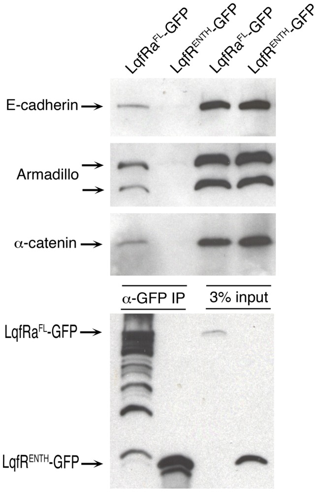
Figure 7. Coimmunoprecipitation of LqfRa and Wingless pathway proteins.

Shown is a blot of protein extracts, before and after immunoprecipitation, from embryos expressing either LqfRaFL-GFP or LqfRaENTH-GFP as a negative control. The LqfR protein fusions were expressed from UAS transgenes using an Actin5C-gal4 driver. The two leftmost lanes (α-GFP IP) are immunoprecipitates using GFP antibodies, and the rightmost lanes (3% input) are aliquots of the protein extracts used, loaded to show that equivalent amounts of protein were present in each extract subjected to immunoprecipitation.
