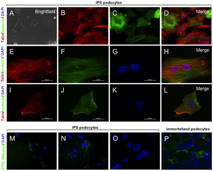
Figure 3. Functional contractility and permeability.
Live cell imaging was used to record the response of iPS podocytes to the addition of AII (See Movie S1). A) Phase contrast imaging of iPS podocytes following the addition of AII at time 0. The iPS podocytes were transduced with RFP-actin (B) and immunostained with the contractile protein, podocin (C). D) Merge image of actin (red) and podocin (green) with DAPI-stained nuclei (blue). E) Confocal immunofluorescence shows that iPS-podocytes transduced with RFP-talin (E) co-expressed podocin (F) at time 0. G) DAPI-stained nuclei and merged image are shown (H). After 6 hours in culture, the iPS podocytes were viable and display a contracted morphology in response to AII (I–L) where RFP-talin (red), podocin (green) and DAPI (blue) are shown. (M-N) By fluorescence microscopy, the iPS podocytes were able to uptake FITC-albumin (green) into the cytoplasm when cultured at 37°C, compared to iPS podocytes cultured at 4°C (O) that served as a control. (P) Immortalized human podocytes also showed endocytosis of FITC-albumin in a similar morphological pattern. Mag A–D ×200; E–L, N-P ×1000; M ×100. Abbreviations: angiotensin II (AII); red fluorescent protein (RFP).

