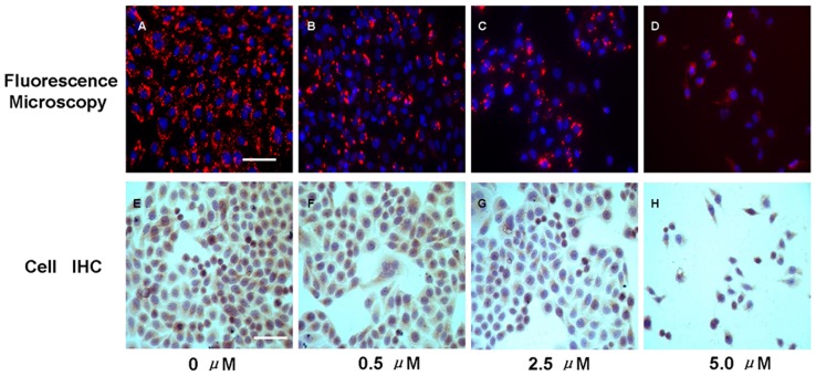
Figure 1. Effects of As2O3 on A431 cell EGFR expression.
Cells were exposed to different concentrations of As2O3. At 48 h post-treatment, cells were assessed by fluorescence microscopy for visualization of the intake of EGF-Cy5.5 and cell immunohistochemistry for assay of EGFR expression. A–D, representative fluorescence images of different groups (Scale bar = 100 µm), Cy5.5 was pseudo-colored red, DAPI was pseudo-colored blue; E-H, representative images of cellular EGFR Immunohistochemistry assay (Scale bar = 100 µm), diaminobenzidine (DAB) showed as brown color represented EGFR expression and hematoxylin showed as blue color indicated the cellular nuclear. Interestingly, cell numbers decreased as the arsenic trioxide concentration increased from 0 µM to 5 µM in both fluorescent and immunostained images, indicating that As2O3 induced a dose-dependent inhibition on tumor cell proliferation as previously reported [31].

