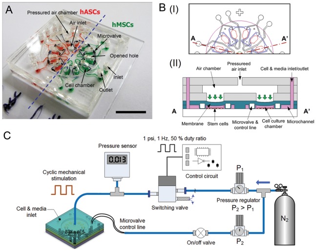
Figure 1. Microchip and experimental setup for evaluating stem cells towards osteogenesis under mechanical stimulation.
(A) The microchip is comprised of a cover, an air chamber, looped microvalves, and twelve cell culture chambers. These paired cell chambers share the inlet/outlet channel. The cells (hMSCs and hASCs) are loaded into half of the chip, individually. Scale bar = 1 cm. (B) Schematic diagram of top view (I) and simplified cross-sectional view (II) of the device. The device was designed to culture two different stem cells simultaneously and to apply mechanical stimulation using cyclic pneumatic force. (C) The experimental setup for mechanical stimulation, including a controlled nitrogen gas pressurized air chamber. The frequency of pneumatic pressure is controlled with a switching solenoid valve derived by a control circuit. During mechanical stimulation, microvalves are closed with higher pressure (P2>P1) to prevent undesired shear stress in the cell chambers.

