
Figure 2. Regions of interest (ROIs) drawn on coregistered MR images.
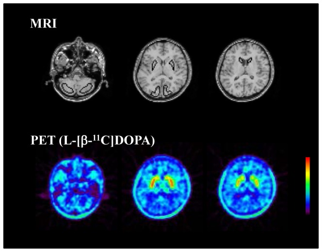
ROIs are defined for the cerebellar cortex, putamen, caudate head, and occipital cortex. Typical PET summation images of frames between 29–89 min after intravenous injection of L-[β-11C]DOPA for baseline study are also shown.