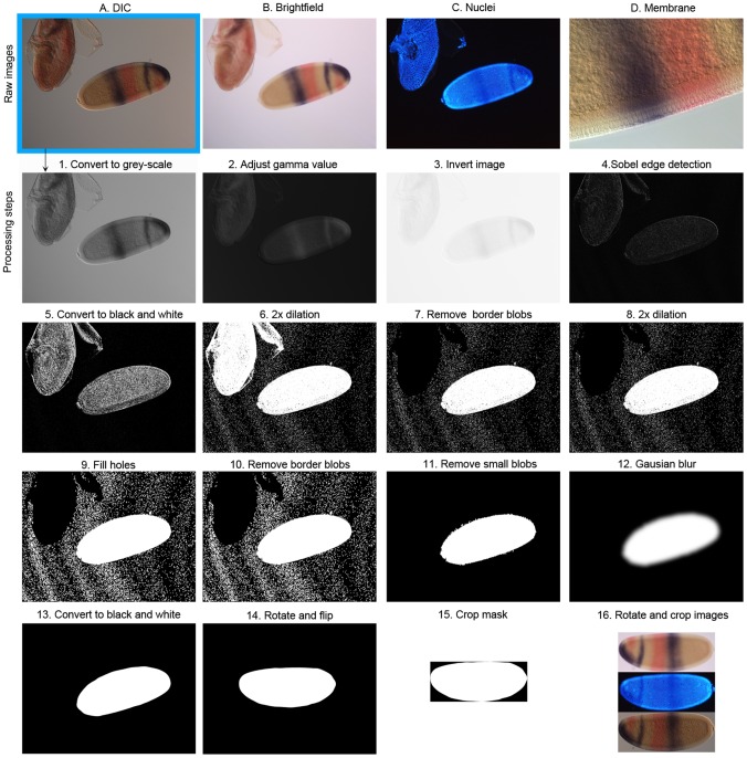
Figure 2. Generating the embryo mask.
The top row displays the four raw images obtained by microscopy: (A) DIC, (B) bright-field, (C) nuclear counterstain, and (D) detailed membrane morphology. The DIC image is used to create the binary embryo mask. This is achieved through a series of processing steps (1–16), which are described in detail in the Materials and Methods section of the main text.

