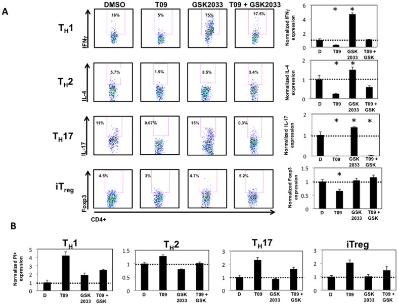
Figure 6. Cell death CD4+ T helper lineages is a function of LXR activity.
A, Intracellular cytokine staining on splenocytes differentiated under TH1, TH2, TH17, or iTreg conditions and treated with vehicle (DMSO), T09 (5 µM), GSK2033 (5 µM), or a combination of T09 and GSK2033 (5 µM each) for the duration of the time course (4 days). Graphs summarizing the FACS plots with data normalized to DMSO controls. B, The effect of T09 (5 µM), GSK2033 (5 µM), or a combination of T09 and GSK2033 (5 µM each) on the viability of TH1, TH2, TH17, and iTreg cells. Cells were FACS analyzed and gated on PI positive cells and normalized to DMSO controls. (n = 4, * p<0.05).

