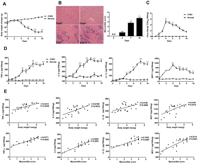
Figure 1. Expression kinetics of pro-inflammatory cytokines and their correlations with the severity of acute myocraditis.
Male BALB/c mice were infected with 103 TCID50 of CVB3 at day 0. (A) The body weight changes were monitored daily until day 10 post-infection. (B) Paraffin sections of heart tissues were prepared on day 0, 4, 7, 10 respectively and cardiac inflammation was revealed by H&E staining (magnification: ×200) (left). The severity of myocarditis was scored by a standard 0–4 scale according to the foci of mononuclear infiltration and myocardial necrosis (right). (C) Hearts were removed aseptically, weighed, and homogenized daily post-infection for TCID50 assay. (D) Hearts were collected and homogenized daily post-infection. The expression of pro-inflammatory cytokines (TNF-α, IL-6, IL-1β and MCP-1) were analyzed by ELISA assay. (E) Correlations between pro-inflammatory cytokines expression levels in cardiac tissues and measures of the severity of acute myocarditis (body weight loss or myocarditis pathological score) at day 7 following 103 TCID50 CVB3 inoculation. Results were presented as the means±SEM of three separate experiments.*, P<0.05; **, P<0.01; ***, P<0.001. Each group contained 8 mice.

