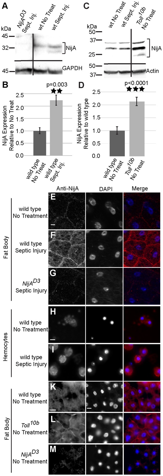
Figure 1. Ninjurin A protein response to septic wounding.

(A) Western blot of whole adult male lysates probed with anti-NijA. NijA increases expression two hours after infection in adults. NijAD3 null lysates demonstrate antibody specificity. Black lines indicate regions of the blot that were omitted for clarity. (B) Graph representing three replicates of the western blot pictured in (A). NijA levels increase significantly in adults after septic injury (p = 0.003). (C) Western blot of whole male larval lysates probed with anti-NijA. NijA levels do not change 2 h after septic injury in third instar larvae; in contrast larval Toll10b gain-of-function mutant larvae have increased levels of NijA protein. (D) Graph representing five replicates of the western blot pictured in (C). NijA levels increase significantly in constitutively activate Toll10b mutant larvae (p<0.0001). (E–M) Anti-NijA (red) and DAPI (blue) labeling nuclei. All scale bars are 10 µm. (E–G) Anti-NijA stained non-permeabilized fat bodies of male third instar larvae show an increase in NijA at the cell surface 2 h after septic injury (compare E and F). (G) NijAD3 larvae demonstrate the NijA antibody specificity. (H,I) Anti-NijA stained non-permeabilized hemocytes of third instar larvae ex-vivo show an increase in NijA at the cell surface 2 h after septic injury. (K–M) Anti-NijA stained permeabilized fat bodies of male third instar larvae show increased NijA expression in gain-of-function Toll10b mutants. Error bars in (B,D) represent standard error of the mean.
