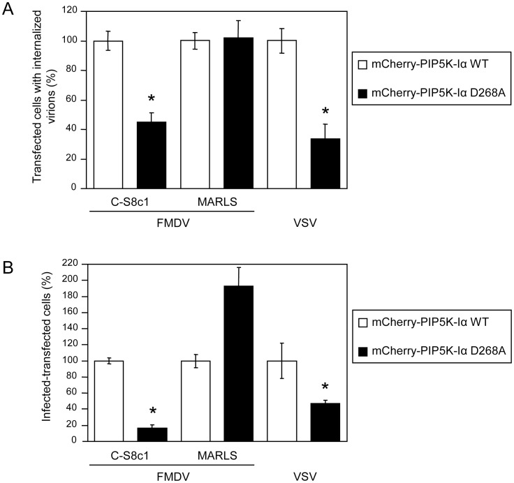
Figure 5. PIP5K-Iα is involved on entry and infection of FMDV C-S8c1 and VSV.
(A) BHK-21 cells transfected with mCherry fused to WT or a KD version of PIP5K-Iα (mCherry-PIP5K-Iα WT and mCherry-PIP5K-Iα D268A, respectively) and 24 h later were incubated with the different FMDV variants (C-S8c1 and MARLS) or VSV (MOI of 70 PFU/cell) for 25 min and processed for immunofluorescence. The graph represents the percentage of cells that showed internalized virus determined as described in Materials and Methods. At least 100 transfected cells per coverslip were scored for each assay (3 coverslip). (B) BHK-21 cells were electroporated with a plasmid encoding mCherry-PIP5K-Iα WT as control, or mCherry-PIP5K-Iα D268A. At 24 h post-electroporation, monolayers were infected with the corresponding virus (MOI of 1 PFU/cell) and cells were fixed and processed for immunofluorescence at 7 h post-infection. Bars represent the mean percentage of transfected and infected cells ± SD, normalized to the level of infection of cells expressing the mCherry-PIP5K-Iα WT. Statistically significant differences between cells transfected with mCherry-PIP5K-Iα WT or D268A are indicated by an asterisk (ANOVA P≤0.05).

