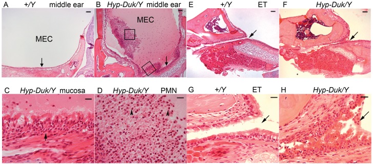
Figure 2. Typical middle ear pathology on H&E sections from Hyp-Duk/Y mice at 12.
weeks of age. (A) Middle ear section shows the clear middle ear cavity (MEC) and normal epithelial layers (indicated by an arrow) in control mice (+/Y). (B) There was a large amount of effusion in the MEC and the middle ear mucosae (indicated by an arrow) were thickened in the Hyp-Duk/Y mice. (C) Enlargement from (B) (lower box) shows hyperplastic ciliated epithelial cells and expanded lamina propria connective tissue in the mucoperiosteum (arrow). (D) Enlarged from (B) (central box) shows the effusion material containing mainly polymorphic nuclear cells (PMN, indicated by arrowheads). Eustachian tubes (ET) as indicated by arrows appear normal in the +/Y mice with a single layer of epithelial cells (E and G; G enlarged from E), but exhibit narrowed and hyperplastic ciliated epithelial cells in the Hyp-Duk/Y mouse (F and H; H enlarged from F). Scale bars in A, B, E, and F: 100 µm; scale bars in C, D, G, and H: 20 µm.

