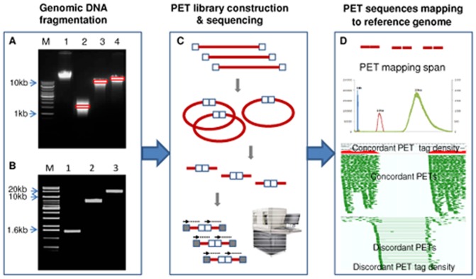
Figure 1. DNA-PET library construction, sequencing and mapping.
(A) The genomic DNA was randomly sheared to different size range. (B) The very narrow region DNA fragments were obtained after size selection. (C) The purified DNA fragments were circularized, EcoP15I digested, sequencing adaptor ligated, and finally sequenced by SOLiD sequencer. (D) PET mapping span distribution of 1 kb (blue), 10 kb (red) and 20 kb (green) libraries. Based on the mapping pattern, PETs can be distinguished as concordant PETs and discordant PETs.

