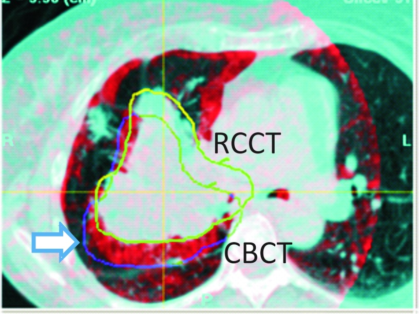
Figure 10.
Overlay of coronal sections from the respiration-correlated CT (RCCT, blue enhanced) and respiration-correlated cone-beam CT (CBCT, red) of Patient 7. The images are aligned to the vertebral column. Yellow curve indicates outline of the GTV drawn on the RCCT; blue curve indicates location in the cone-beam CT of the GTV, which has shifted 14 mm posteriorly relative to the RCCT (arrow).

