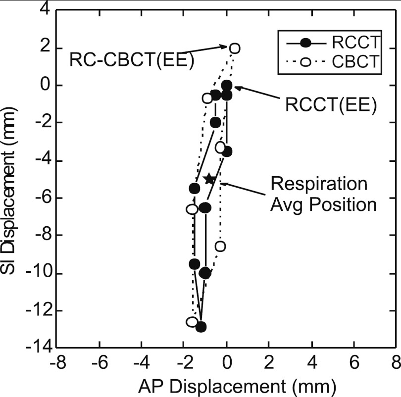
Figure 2.
Example tumor trajectories in the anterior-posterior (A/P) and superior-inferior (S/I) directions, obtained from the respiration-correlated CT (RCCT) and one of the respiration-correlated cone-beam CT (RC-CBCT) image sets of Patient 11. Each point indicates displacement relative to the 50% phase reference point (approximately end expiration, EE) in the RCCT. Star symbol indicates the respiration average position in both image sets.

