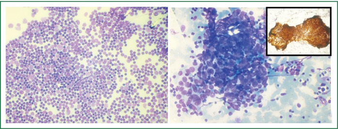
Figure 3.
A. Fine needle aspirate of mediastinal mass showing numerous clusters of epithelial cells displaying mild to moderate atypia in a background of abundant reactive lymphoid tissue (May-Grϋnwald-Giemsa stain × 200); B. Fine needle aspirate from pleural based lesion showing similar morphology confirming metastatic thymic carcinoma (May-Grϋnwald-Giemsa stain × 400); Inset shows strong cytokeratin immunoreactivity in the thymic epithelial cells (Immunoperoxidase with Envision).

