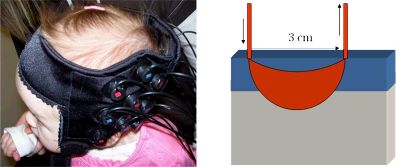
Figure 1.
Left: Infant wearing a cap holding an array of 9 optical fibers to gather data from 12 NIRS channels over the left temporal cortex. Right: Schematic of a NIRS channel, showing the input and output optical fibers and the banana-shaped pathway of photons that dip into the gray matter of the brain after passing through superficial layers of skin, skull, and surface vasculature.

