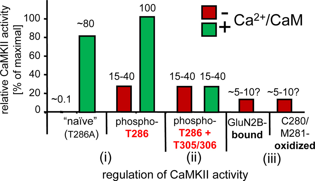
Figure 2.
Levels of CaMKII activity in response to different forms of regulation. (i) Naïve CaMKII shows only low basal activity (~0.1% of maximum), but is fully activated by Ca2+/CaM-stimulation [2]. This is accompanied by rapid T286-autophosphorylation; when T286-autophosphorylation is prevented by T286A mutation, the activity level remains slightly lower [58]. After T286-autophosphorylation, CaMKII remains partially active even after dissociation of Ca2+/CaM (autonomous), but this autonomous activity is significantly further stimulated by Ca2+/CaM [29]. (ii) T305/306-autophosphorylation of T286-phosphorylated CaMKII prevents Ca2+/CaM binding and thus further stimulation of the autonomous activity. (iii) Autonomous activity can also be induced by GluN2B binding [61] or by C280/M281 oxidation [30]; like T286-autophosphorylation, both mechanisms require an initial Ca2+/CaM-stimulus [30, 61]. The levels of “autonomy” (the ratio of autonomous over maximal CaM-stimulated activity) after GluN2B binding or oxidation are estimated adjustments (based on conditions in the original reports that overestimate autonomy, either by use of an autonomy-favoring substrate [29, 61] or by conditions resulting in unusually low rates of stimulated activity [30]). The activity estimates shown are based on an adapted compilation from [29, 30, 58, 61].

