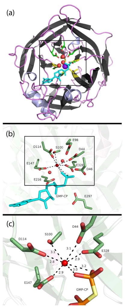
Figure 1.
(a) Cartoon depiction of the hSCAN-1 5-bladed beta-propellar crystal structure (PDB code: 1S1D).4 The crystalized structural calcium is centered in purple, and the five calcium-coordinated carbonyl groups (one from each beta sheet) are highlighted in yellow. Three crystalized waters in the active site are shown as red spheres, proximal acidic ionizable residues are shown as sticks, and the substrate analog, GMP-CP, is depicted in cyan. (b) A closer view highlighting the eight ionizable aspartates and glutamates and a serine, all within 5.5 Å of the negatively charged phosphates. (c) A close-up of the apparent hepta-coordination around a crystal-assigned water, or potentially misassigned cation. The distances are shown in Å.

