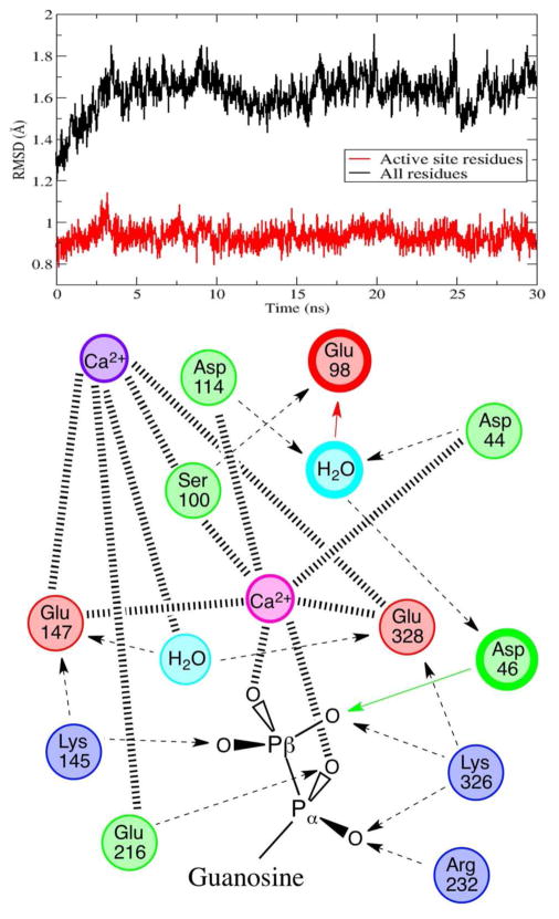
Figure 3.
Top: heavy atom RMSD from the crystal structure for all residues within 8 Å of the terminal phosphate (in red), highlighting active site stability for 30 ns of molecular dynamics, and for the entire enzyme (in black). Bottom: schematic of the structural interactions between the active site residues, the structural calcium, the catalytic calcium, the nucleophilic water, an active site water, and the GDP substrate. Hashed lines represent coordination to calcium, dashed lines represent hydrogen bonding with arrows pointing to H-bond acceptors, colored arrows and bold circles depict proton transfers and residues involved in the catalytic chemistry.

