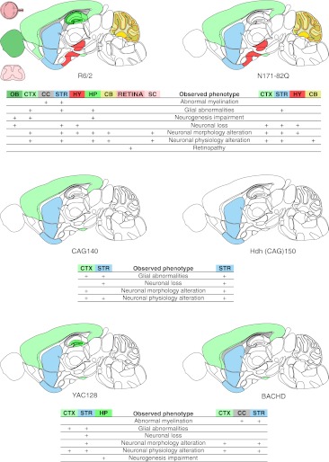
Fig. 2.
Diagrams of neuropathology in the brain, spinal cord, and retina of selected mouse models of Huntington’s disease. Seven neuropathology phenotypes, namely, neuronal loss, neuronal morphology alterations, neuronal physiology alterations, glia abnormalities, retinopathy, neurogenesis impairment, and abnormal myelination, were selected, along with the localization available for these phenotypes in CNS region column of the data table. The brain regions involved in the selected neuropathology phenotypes for R6/2, N171-82Q, CAG140, Hdh(CAG)150, YAC128, and BACHD models were marked with colors on a schematic sagittal section of the mouse brain. Brain regions were color-coded using colors from the Allen Brain Atlas [374]. The main brain regions involved in the HD models are the striatum (STR) and cerebral cortex (CTX). The R6/2 and YAC128 models also show involvement of the hippocampus (HP) as a region of “neurogenesis impairment” [375, 376]; the deficit is also observed in the piriform cortex (CTX, bottom part) [377] and olfactory bulb (OB) [378] of the R6/2 mice. Moreover, R6/2 neuropathology is observed in the hypothalamus (HY), cerebellum (CB), spinal cord (SC), and retina of the eye (RETINA). The cerebral cortex (CTX) is not involved in the selected neuropathology phenotypes in the Hdh(CAG)150 knock-in. As in R6/2 animals, abnormal myelination has been detected in the corpus callosum (CC) and striatum (STR) of BACHD animals

