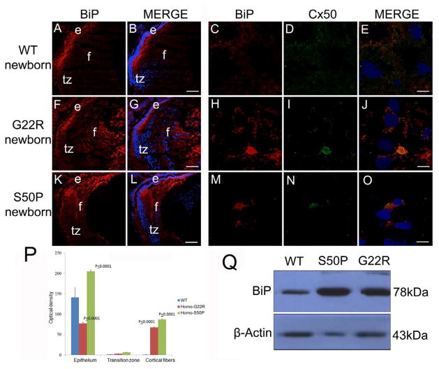
Figure 4.
BiP levels are elevated in newborn Cx50 mutant lenses (A, B,) Wildtype newborn lenses express BiP prominently in the lens epithelium with lower amounts in the lens fibers that have not completed organelle degradation. (C,D,E)- BiP and Cx50 are not found co-localized in wildtype lens fibers at birth (F, G,) Homozygous Cx50G22R lenses have increased levels of BiP expression, particularly in the lens fiber cells. (H,I,J)- The elevated amounts of BiP found in G22R lens fibers is co-localized with Cx50 (K,L) Cx50S50P lenses also have increased levels of BiP in the lens fibers although this increase is more confined to the cortical fiber cells and this additional BiP co-localizes with Cx50 (M,N,O). (P) Quantitation of BiP levels in G22R/G22R and S50P/S50P homozygotes in the lens epithelium, transition zone and cortical fibers using ImageJ. P values reported represent ANOVA analysis performed on results obtained from at least three biological replicates. (Q) Western blotting analysis of newborn lenses confirming the increase in BiP expression in the Cx50S50P and Cx50G22R mutants. Red-BiP; Blue-DRAQ5(DNA); Green- Cx50 e- epithelial cells; f- fiber cells; tz-transition zone; Scale bar A,B,F,G,K,L=77 μm, C,D,E,H,I,J,M,N,O= 6 μm

