Figure 4.
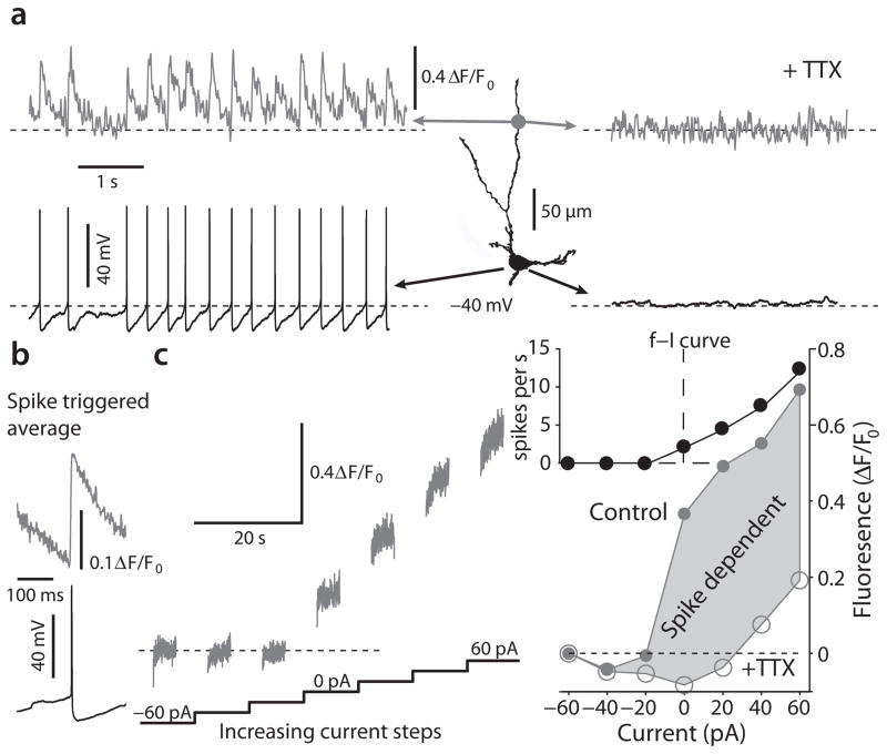
2PLSM calcium imaging demonstrates that calcium dynamics are dominated by spike associated influx. (a) Left: Somatic voltage recording and 2PLSM Fluo-4 imaging from a distal dendritic location (140 μm from soma) of a DMV cell reveal spontaneous discharge that is accompanied by spike associated calcium transients. Right: Application of TTX reveals a stable resting potential without any subthreshold calcium oscillations. (b) spike triggered average of calcium transients in a DMV neuron reveal a rapid increase in calcium that is followed by an exponential decay. (c) Left: 2PLSM calcium imaging from the soma in response to a sequence of 10-s current injections from −60 pA to +60 pA to either hyperpolarize and silence discharge or to depolarize and increase discharge. The value of fluorescence for 0 pA is defined as the ambient fluorescence. Right: bottom – the plot of fluorescence as a function of current before (filled circles) and after (empty circles) TTX treatment demonstrates that the majority of calcium entry is spike-dependent. At the population level, the rise in free calcium as a function of applied current, estimated from the slope of this function from −20 to +60 pA, decreased significantly in TTX (not shown, n = 6 cells, P < 0.05, SRT). top – frequency-intensity (f–I) curve of this neuron in response to the same current steps.

