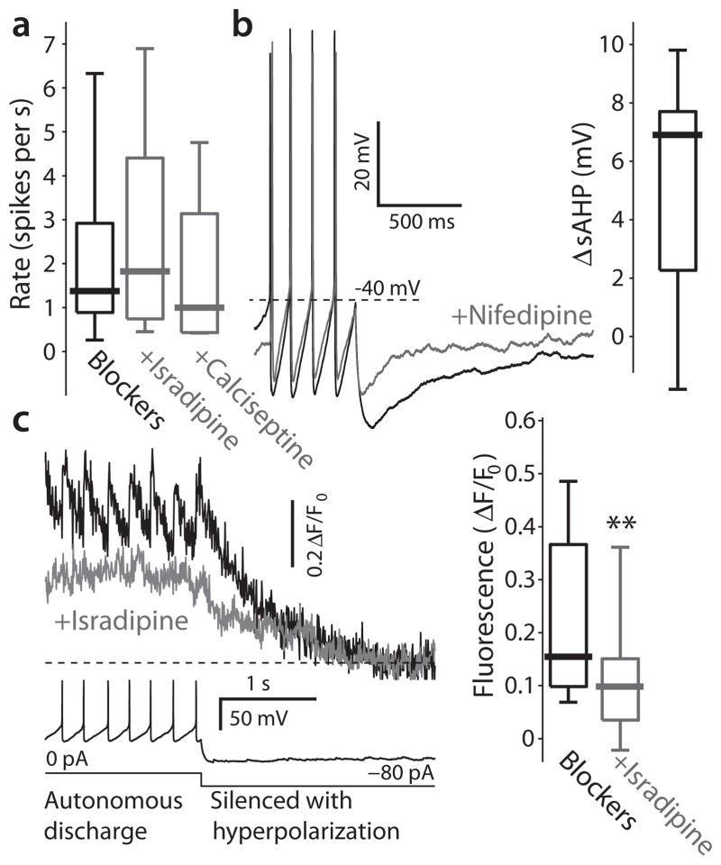
Figure 5.
Contribution of Cav1 channels to discharge patterns and ambient calcium levels in DMV neurons. (a) Preincubation in 200 nM of either isradipine or calciseptine, that antagonize Cav1 channels, does not alter the distribution of DMV neurons firing rates. (b) Application of 5 μM nifedipine, a Cav1 channel antagonist, significantly reduced the depth of the slow afterhyperpolarization (sAHP), measured from spike threshold, that follows long depolarizing pulses. Inset: distribution of changes in sAHP amplitude (ΔsAHP) in response to this drug. (c) Left: recording of spontaneous discharge followed by a hyperpolarizing pulse to silence the cell (bottom) can be used to measure the ambient fluorescence during autonomous discharge (top). Treatment with 5 μM isradipine consistently reduces the baseline level of fluorescence in cholinergic DMV neurons. Right: distribution of ambient levels of fluorescence before and after treatment with 5 μM isradipine ** P < 0.01, SRT.

