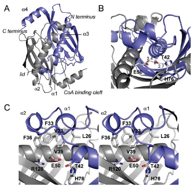
Figure 3.
Crystal structure of FlK. (A) Cartoon representation of the FlK crystal structure looking down at the active site and lid structure formed by α1 and α2 (residues 23–26 and 31–34, repectively). Chain A is colored in blue, and chain B is colored in gray (α3, residues 42–58; α4, residues 125–135). (B) View of the hydrogen- bonding network at the putative active site showing Thr 42, His 76, and Glu 50 site water. (C) Stereoview of the active site and the hydrophobic lid (carbon, gray; nitrogen, blue; oxygen, red; sulfur, yellow).

