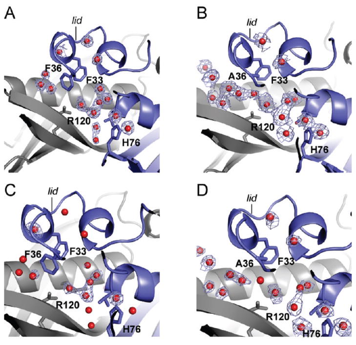
Figure 5.
Accessibility of the active site to water in FlK and FlK-F36A. Electron density maps are shown in light blue, chain A is colored in blue, chain B is colored in gray, and water molecules are shown as red spheres. (A) 2Fo − Fc map contoured at 0.8σ for water molecules at the active site of wild-type FlK. (B) 2Fo − Fc map contoured at 0.8σ for water molecules at the active site of FlK-F36A. (C) Fo − Fc solvent omit map contoured at 2.5σ for wild-type FlK overlaid with the refined FlK structure. (D) Fo − Fc solvent omit map contoured at 2.5σ for FlK-F36A overlaid with the refined FlK-F36A structure.

