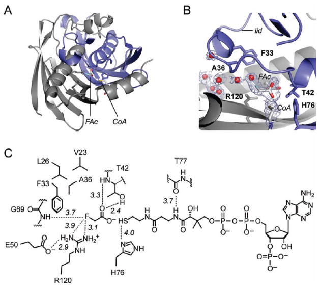
Figure 6.
FlK-F36A product complex. Chain A is colored in blue, and chain B is colored in gray. For clarity, only the fluoroacetate and CoA ligands are colored by atom (carbon, gray; nitrogen, blue; oxygen, red; fluorine, light green; sulfur, yellow). (A) A view of the product bound at the FlK-F36A active site shown in the context of the overall fold. (B) Fo − Fc solvent/ligand omit map contoured at 2.5σ overlaid with the refined FlK-F36A product complex structure. (C) Chem Draw figure showing interactions observed between FlK-F36A and the products.

