
FIGURE 11.
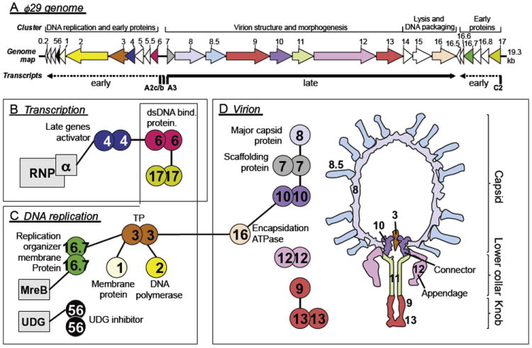
Genome map and literature curated ϕ29 interactome. (A) Genome and simplified transcriptional map of ϕ29 (after Meijer et al., 2001a). Major promoters are indicated by their names, and major transcripts are shown as dashed (early) and solid (late) arrows. Other promoters are not indicated for clarity. Open reading frame (ORF) arrows indicate transcription direction. The color of ORF symbols corresponds to the color used for protein symbols in B, C, and D. PPIs known for ϕ29, organized by their cellular function (transcription, DNA replication, or virion). B. subtilis proteins are shown as rectangles, phage proteins as circles. Equal protein symbols contacting each other indicate homomers. Heteromeric PPIs are highlighted by protein symbols that are connected by lines. (D) Schematic cross view of the mature ϕ29 virion (after Xiang et al., 2008). UDG, uracil–DNA glycosylase; RNAP, RNA polymerase; TP, terminal protein.