
FIGURE 12.
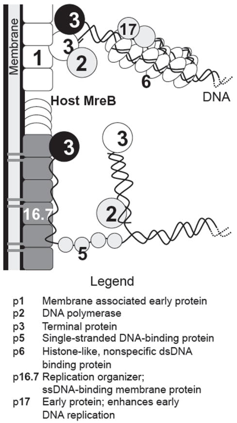
Interactions in ϕ29 genome replication and membrane localization. Proteins that contact each other have been shown to interact. For clarity, only one genome end is shown. Parental TP is highlighted by a black symbol, whereas primer TP is in white. The upper part of the figure includes PPIs that preferentially play a role in replication initiation. The lower part of the figure indicates elongated DNA with the associated ssDNA regions. Because p16.7 and p5 (SSB) both bind ssDNA, they could either redirect the parental ssDNA strand to the membrane via TP-p16.7 and p16.7–ssDNA interactions or stabilize it in the cytoplasm via SSB alone (Meijer et al., 2001b). After Bravo et al. (2000) and Serna-Rico et al. (2003).